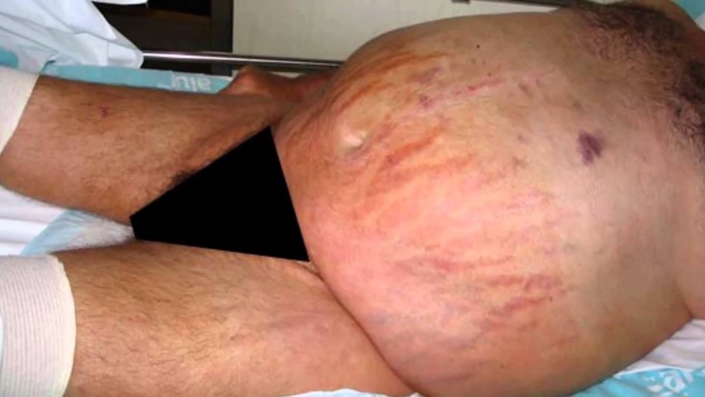
Cushing’s disease is a cause of Cushing’s syndrome characterised by increased secretion of adrenocorticotropic hormone (ACTH) from the anterior pituitary (secondary hypercortisolism). This is most often as a result of a pituitary adenoma (specifically pituitary basophilism).
Symptoms
The symptoms of Cushing’s disease are similar to those of Cushing’s syndrome: patients with Cushing’s disease usually present with one or more signs and symptoms secondary to the presence of excess cortisol
Symptoms include:
- moon face (resulting in a large chin and large forehead)
- extra fat around neck
- easy bruising of the skin
- purplish stretch marks on abdomen
- weight gain
- red stretch marks
- excess hair growth (women)
- fatigue
- diabetes mellitus (less common)
- irregular menstruation
- high blood pressure
- irritability
Diagnosis
Diagnosis is made first by diagnosing Cushing’s Syndrome, which can be difficult to do clinically since the most characteristic symptoms only occur in a minority of patients. Some of the biochemical diagnostic tests used include salivary and blood serum cortisol testing, 24-hour urinary free cortisol (UFC) testing, the dexamethasone suppression test (DST), and bilateral inferior petrosal sinus sampling (BIPSS).
No single test is perfect and multiple tests should always be used to achieve a proper diagnosis. Diagnosing Cushing’s disease is a multidisciplinary process involving
- doctors
- endocrinologists
- radiologists
- surgeons
- chemical pathologists
After a diagnosis is done an MRI can determine if a pituitary tumor is visible.
In Cushing’s disease, typically very small (microadenoma) tumors are found. If no tumor is visible, then inferior petrosal sinus sampling is indicated.

Hormone Testing
The testing for excessive cortisol levels can be complex and challenging. Blood tests may not detect the presence of excessive cortisol secretion because blood levels of cortisol naturally vary throughout the day. Therefore a simple measurement of the blood cortisol level is usually not definitive. Tests we will order include:
- 24-hour urine free cortisol (UFC) measurement
- Cortisol saliva testing
- Blood ACTH
- If low, may suggest the presence of an adrenal tumor producing cortisol
24-hour urine free cortisol (UFC) measurement
The amount of cortisol found in urine collected over a 24-hour period is an important measurement in diagnosing Cushing’s syndrome. This test sums the total daily production of cortisol.
- In Cushing’s disease, the 24-hour urine free cortisol (UFC) levels are typically at least four to five times greater than normal.
- Your doctor may need two or more elevated 24-hour UFC tests to confirm the diagnose Cushing’s syndrome.
- Uncommonly, Cushing’s disease can come and go (“periodic Cushing’s disease”), requiring multiple 24-hour UFC tests.
- Cortisol saliva testing
In some cases, an endocrinologist may request testing for the level of cortisol in the saliva. Similar to blood cortisol levels, the amount of cortisol in the saliva at or near midnight is expected normally to be very low. Multiple elevated midnight salivary cortisol levels may help confirm the diagnosis of Cushing’s syndrome.
It is important not brush the teeth just prior to collecting the sample.
This test may not be valid in smokers.
MRI Imaging
Once your doctor suspects Cushing’s disease based on clinical findings and hormonal testing, a Magnetic Resonance Imaging (MRI) scan of the pituitary gland is the best way to detect the presence of an adenoma in Cushing’s disease.
MRI detects a pituitary adenoma in about 70 percent of cases. It is important that you undergo these imaging tests at a Pituitary center with special expertise in imaging small tumors.
Inferior petrosal sinus sampling
In some cases, MRI fails to identify an abnormality. If your doctor suspects Cushing’s disease, he or she may order super-selective inferior petrosal sinus sampling (IPSS).
IPSS must be performed by an experienced interventional neuroradiologist because it carries risk, and if done inappropriately, can give misleading results.
The inferior petrosal sinuses are veins that occur on both sides of the pituitary gland.
- Blood leaving the right half of the pituitary gland drains into the right inferior petrosal sinus and vice versa.
- Specially trained interventional neuroradiologists can thread a tiny catheter into the inferior petrosal sinus. Blood samples are then taken from each sinus and from a vein just below the heart.
If the ACTH level is the same in the inferior petrosal sinus compared to the vein below the heart, this suggests that a tumor somewhere else in the body (ectopic, not pituitary) is producing ACTH.
In Cushing’s disease, the ACTH level in the inferior petrosal sinus is much higher compared to the vein below the heart
Treatment Options
Treating Cushing’s disease requires an experienced team of experts.
Treatment options include:
- Surgery
- Medication
- Radiation therapy
- Other treatment options
Surgery
The first-line treatment of Cushing’s disease is surgical resection of ACTH-secreting pituitary adenoma; this surgery involves removal of the tumor via transsphenoidal surgery (TSS).
• When an experienced pituitary surgeon performs the operation, the cure rate for smaller tumors (microadenomas) is 80 percent to 85 percent. If the tumor spread into nearby internal structures, the cure rate is 50 percent to 55 percent.
• Neurosurgeons who specialize in pituitary tumor surgery utilize a technique that removes the tumor as one piece. They find the tumor and dissect it around the edge.
• The endoscopic endonasal approach is a minimally invasive approach, using your natural nasal passageway. It does not require a head incision.
• An endoscopic technique can be very effective in safely removing tumor, while at the same time minimizing hospitalization time and discomfort.
Radiation Therapy for Cushing’s Disease
In some cases, surgeons may not be able to remove the tumor surgically. Radiation therapy can be very effective in controlling the growth of these tumors.
Stereotactic radiosurgery is a technique in which a highly focused of radiation can be delivered to the tumor target. Because the radiation beam is carefully sculpted, the surrounding brain structures receive only a fraction of the radiation dose and are typically unharmed (with the exception of the normal pituitary gland).
A consequence of radiation treatment is that it can cause delayed pituitary failure. This typically occurs several years after treatment, and therefore continued long-term follow-up with an endocrinologist is important. Hormone replacement may be required.
 English
 Italiano
![cushings-hisuitism[2]](http://www.ipofisi.com/wp-content/uploads/2016/03/cushings-hisuitism2.jpg)